Mammography
An estimated one in every eight women will develop breast cancer sometime in her life. Breast cancer is the second-highest cause of cancer death in women, second only to lung cancer.
Mammography is an invaluable early detection tool for breast cancer. A baseline mammogram is recommended for women ages 35 to 39. Women 40 and older should have a mammogram every year.
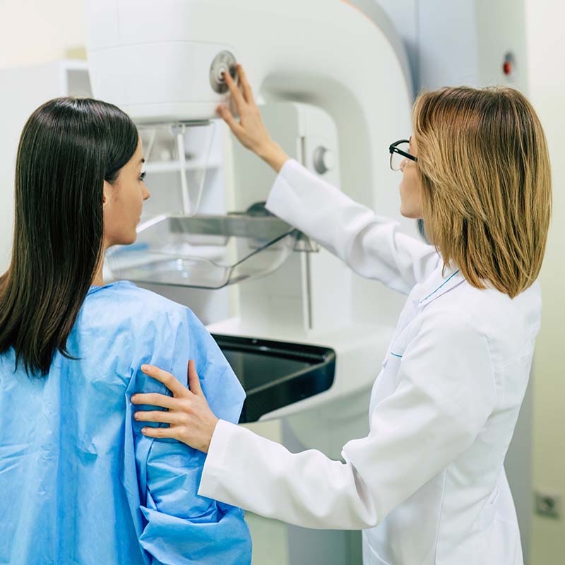
At OB/GYN Associates of Alabama, we want to make your mammogram as comfortable as possible, both physically and mentally. We offer a highly experienced technician who provides a thorough 3D screening and the latest educational information on breast care.
What is a 3D Mammogram?
Our team at OB/GYN Associates offers 3D mammography, providing a clearer and more accurate view of the breast. Also known as breast tomosynthesis, a 3D mammogram uses imaging with various X-rays to test breast tissue. By taking multiple images of the breast, our doctors clearly view multiple tissue layers and any potential problems.
When you get a 3D mammogram to screen for breast cancer, you’ll receive both the standard 2D imaging procedure, as well as the 3D one because both tests are useful to find breast cancer precursors and other breast abnormalities.
Benefits of a 3D Mammogram:
Mammograms are a great tool to detect breast cancer before lumps appear or symptoms arise. A 3D mammogram has many additional benefits to a patient, such as:
Mammogram FAQs
Every medical screening involves certain risk factors. The ones for a 3D mammogram are as follows:
Radiation exposure. We’re talking low radiation levels with any kind of mammogram because technicians use X-rays to take pictures of a woman’s breasts. It’s important for a woman to know this because a 3D mammogram involves slightly higher levels of radiation exposure than standard imaging simply because the 3D version combines 3D technology with 2D technology.
There are newer 3D mammogram imaging machines that can take 3D and 2D pictures simultaneously, which is believed to limit radiation exposure.
3D mammograms may detect something else entirely. It’s certainly possible that by having a 3D mammogram done, your doctor finds something abnormal. You’ll then need further testing, which may find that your abnormality was benign all along. This is called a false-positive. With a false-positive result, you’re likely to suffer unnecessary anxiety, which is never fun and might lower your quality of life as you wait for the results to come in.
Not all forms of cancer are found. Sometimes, a 3D mammogram fails to detect a specific cancer area, especially if the site is minute or in an obscure place.
You’ll want to do the following as prep for a 3D mammogram:
- Choose carefully where you’d like to have your 3D mammogram done. While this kind of mammogram is becoming increasingly more popular, it’s still not offered everywhere. If this is a procedure you think you’d like, inquire with our front desk and schedule an appointment.
- Make sure insurance covers the procedure. Your particular insurance may not cover a 3D mammogram. Make sure to check with the provider to see what you might need to pay out of pocket. Do this before the procedure so that you’ll not have any unwanted or costly surprises. You may have to pay for the 3D portion of the imaging while your insurance covers the standard imaging test.
- Get the 3D mammogram done when your breasts are tender. Chances are, your breasts are more tender during PMS. Schedule your 3D mammogram during this time for better results.
- Bring any mammogram images you’ve already had done to your appointment. You might be visiting a new medical facility for your 3D mammogram. If this is the case, bring mammogram images you might have from the past so that your new doctor can compare new and old images.
- Refrain from putting on deodorant before having a mammogram. The metallic particles that lurk within various antiperspirants, deodorants, lotions, and perfumes interfere with imaging results. Don’t put anything topical on your bosoms or underarms before a mammogram.
Once you arrive at our offices, you’ll need to prepare. This simply means removing jewelry and clothing from the torso. You’ll wear a gown over your torso during the actual procedure. You’ll probably want to wear a shirt and jeans for easier undressing.
During the imaging test, expect to stand directly in front of the imaging machine. Your technician will then put one breast on the platform of the x-ray machine. You’ll have your head positioned, as well as your torso and arms. This positioning is only to ensure a clear view of the breast being examined.
A plastic plate presses the breast up against the imaging platform. You’ll feel pressure on the breast, which spreads the tissue out. Some women feel pain or discomfort during this process. Be sure to let the technician know when enough’s enough.
The 3D X-ray machine moves from side to side, directly above you as it takes images of the breast. You’ll want to stand completely still during this time, and you may even want to hold your breath if it helps.
Releasing pressure from the breast, the x-ray machine repositions for further imaging. Once again, the breast is pressed up against the x-ray platform by the plastic plate. More pictures ensue. Once all this is done, the same procedure occurs with the opposite breast.
A 3-D X-ray machine collects images of the breasts, then computer-synthesized to create a 3D image. The 3D images are then examined to see every detail. 2D images are also created for additional screening.
A radiologist who is a specialist in this area examines all pictures. They are doing so to find abnormalities in tissue that could point to breast cancer. If they think anything looks unusual, your standard mammogram, as well as any images you’ve had done in the past are scrutinized. After all the information is taken into account, the specialist determines whether or not any additional tests are necessary. These can come in the form of MRIs, ultrasounds, or biopsies. Biopsies are taken to test suspicious cells in a lab. This is called pathology testing.
If you have more questions about 3D mammography, please feel free to call us or tap the button below to set up an appointment with our friendly staff.
Request Your 3D Mammogram Today!
Contact Us To Schedule Your Free Consultation

